The cerebellum plays a very important role in the control of posture and voluntary movements. It unconsciously influences the smooth contraction of voluntary muscles and carefully coordinates their actions, together with the relaxation of their antagonists. The cerebellum has no direct pathway to the lower motor neurons but exerts its control via the cerebral cortex and the brainstem.
The gray matter of the cortex throughout its extent has a uniform structure. It may be divided into three layers: (1) an external layer, the molecular layer; (2) a middle layer, the Purkinje cell layer; and (3) an internal layer, the granular layer.
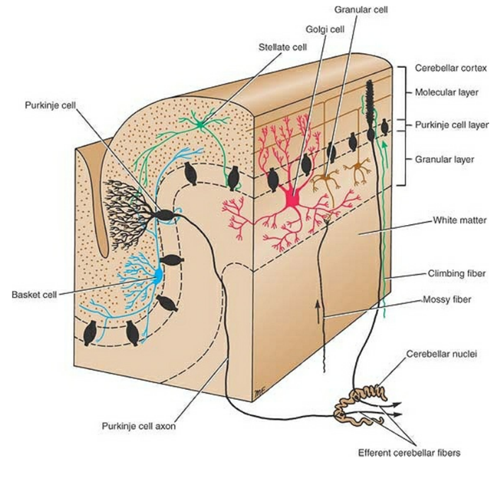
Molecular Layer
The molecular layer contains two types of neurons: the outer stellate cell and the inner basket cell.
Purkinje Cell Layer
The Purkinje cells are large Golgi type I neurons. They are flask shaped and are arranged in a single layer. In a plane
transverse to the folium, the dendrites of these cells are seen to pass into the molecular layer, where they undergo profuse branching. At the base of the Purkinje cell, the axon arises and passes through the granular layer to enter the white matter. On entering the whitematter, the axon acquires a myelin sheath, and it terminates by synapsing with cells of one of the intracerebellar nuclei.
Granular Layer
The granular layer is packed with small cells with densely staining nuclei and scanty cytoplasm. Each cell gives rise to four or five dendrites, which make claw like endings and have synaptic contact with mossy fiber input. The axon of each granule cell passes into the molecular layer, where it bifurcates at a T junction, the branches running parallel to the long axis of the cerebellar folium. These fibers, known as parallel fibers, run at right angles to the dendritic processes of the Purkinje cells. Most of the parallel fibers make synaptic contacts with the spinous processes of the dendrites of the Purkinje cells.
The cortex
The cortex of the vermis influences the movements of the long axis of the body, namely, the neck, the shoulders, the thorax, the abdomen, and the hips. Immediately lateral to the vermis is a so-called intermediate zone of the cerebellar hemisphere. This area control the muscles of the distal parts of the limbs, especially the hands and feet.. The lateral zone of each cerebellar hemisphere appears to be concerned with the planning of sequential movements of the entire body and is involved with the conscious assessment of movement errors.
Intracerebellar Nuclei
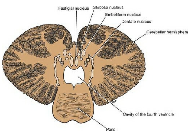
Four masses of gray matter are embedded in the white matter of the cerebellum on each side of the midline. From lateral tomedial, these nuclei are the dentate, the emboliform, the globose, and the fastigial.
The dentate nucleus is the largest of the cerebellar nuclei. It has the shape of a crumpled bag with the opening facing medially. The interior of the bag is filled with white matter made up of efferent fibers that leave the nucleus through the opening to form a large part of the superior cerebellar peduncle.
The emboliform nucleus is ovoid and is situated medial to the dentate nucleus, partially covering its hilus.
The globose nucleus consists of one or more rounded cell groups that lie medial to the emboliform nucleus.
The fastigial nucleus lies near the midline in the vermis and close to the roof of the fourth ventricle; it is larger than the globose nucleus.
The intracerebellar nuclei are composed of large, multipolar neurons with simple branching dendrites. The axons form the cerebellar outflow in the superior and inferior cerebellar peduncles.
Cerebellar Cortical Mechanisms
Certain basic mechanisms have been attributed to the cerebellarcortex.
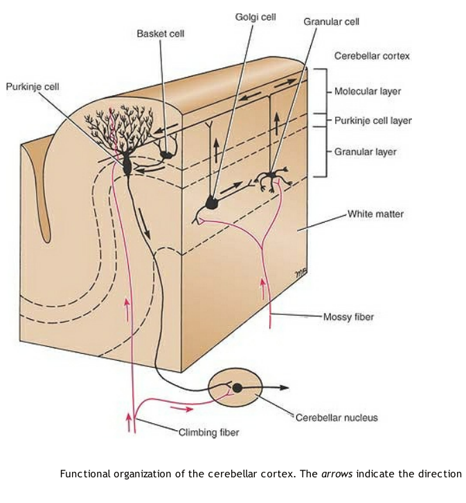
The climbing and the mossy fibers constitute the two main lines of input to the cortex and are excitatory to the Purkinje cells. The climbing fibers are the terminal fibers of the olivocerebellar tracts. They are so named because they ascend through the layers of the cortex. The mossy fibers are the terminal fibers of all other cerebellar afferent tracts. They have multiple branches and exert a much more diffuse excitatory effect. A single mossy fiber may stimulate thousands of Purkinje cells through the granule cells.
Stellate, Basket, and Golgi cells – they serve as inhibitory interneurons. They not only limit the area of cortex excited but influence the degree of Purkinje cell excitation produced by the climbing and mossy fiber input. By this means, fluctuating inhibitory impulses are transmitted by the Purkinje cells to the intracerebellar nuclei, which, in turn, modify muscular activity through the motor control areas of the brainstem and cerebral cortex. It is thus seen that the Purkinje cells form the center of a functional unit of the cerebellar cortex.
Intracerebellar Nuclear Mechanisms
The deep cerebellar nuclei receive afferent nervous information from two sources: (1) the inhibitory axons from the Purkinje cells of the overlying cortex and (2) the excitatory axons that are branches of the afferent climbing and mossy fibers that are passing to the overlying cortex. In this manner, a given sensory input to the cerebellum sends excitatory information to the nuclei, which a short time later receive cortical processed inhibitory information from the Purkinje cells.
Efferent information from the deep cerebellar nuclei leaves the cerebellum to be distributed to the remainder of the brain and spinal cord.
Cerebellar Cortical Neurotransmitters
The excitatory climbing and mossy afferent fibers use glutamate (gamma-aminobutyric acid [GABA]) as the excitatory transmitter on the dendrites of the Purkinje cells. Other afferent fibers entering the cortex liberate norepinephrine and serotonin at their endings that possibly modify the action of the glutamate on the Purkinje cells.