Topic: Applied anatomy of joints of hip and knee
Question: Write a short note on the anatomy of knee joint and its structural components.
Click here for Reference Material-This material is informational alone and is not specifically prepared as an answer for any question. Readers may do their own research before finalising diagnoses according to the characteristics unique to each question. Readers should not proceed without cross-referencing with relevant textbooks as well as standard guidelines available.
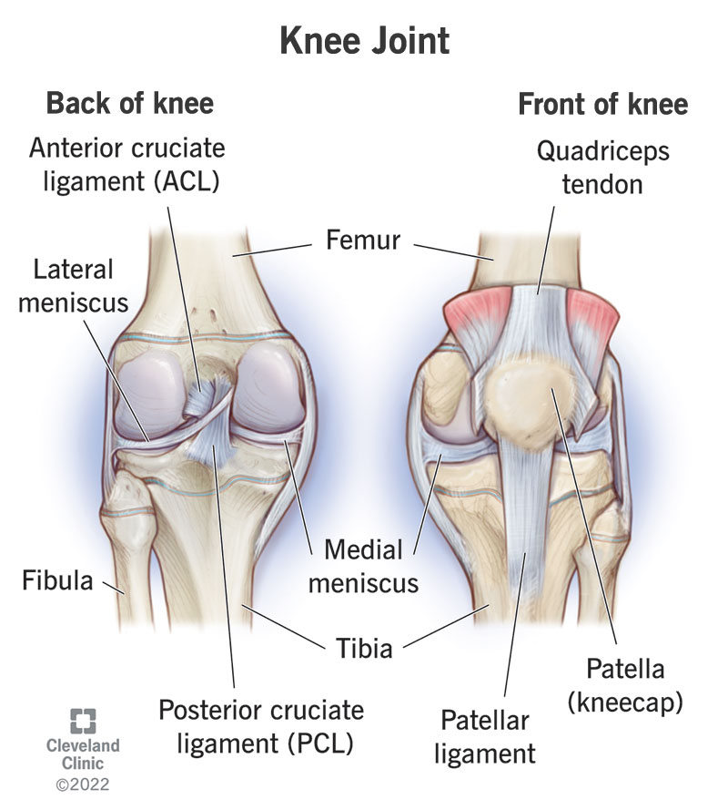
The knee joint is the largest joint in the body. It is a synovial hinge joint that allows flexion, extension and slight internal/external rotation of the leg.The main components of the knee joint include:
• Femur: The distal end of the femur articulates with the tibia to form the knee joint. The femoral condyles are rounded prominences on the distal femur that articulate with the tibial condyles.
• Tibia: The proximal end of the tibia articulates with the femur. The tibial condyles are concave surfaces that articulate with the femoral condyles. The tibia also articulates with the patella anteriorly.
• Patella: The patella is a sesamoid bone embedded in the tendon of the quadriceps femoris muscle. It articulates with the distal femur and acts to protect the knee joint and increase leverage of the quadriceps.
• Articular cartilage: Covers the articulating surfaces of the femur, tibia and patella. It is a smooth, white hyaline cartilage that absorbs shock and allows smooth movement between the bones.
• Synovial membrane: Lines the joint capsule and secretes synovial fluid to lubricate the joint.
• Anterior and posterior cruciate ligaments: Cross within the joint space to connect the femur and tibia and provide anterior/posterior knee stability.
• Medial and lateral collateral ligaments: Connect the medial and lateral aspects of the femur and tibia to provide valgus/varus stability to the knee.
• Menisci: Two C-shaped fibrocartilage pads that sit between the femoral and tibial condyles to provide shock absorption, joint stability and congruity between the otherwise incongruent surfaces.
• Tendons: The quadriceps, hamstring and gastrocnemius tendons all cross the knee joint and facilitate leg movement/joint stabilization.
• Bursae: Fluid-filled sacs that prevent friction from tendons/ligaments over the joint. Prepatellar bursa lies anterior to the patella, while anserine bursae sit medially.
The knee joint’s anatomical complexity gives it a remarkable range of motion and function. Damage or deterioration of any of these structures can disrupt mobility, joint integrity and quality of life. Understanding knee joint anatomy is fundamental to assessment and treatment of knee injuries or degenerative conditions.