Topic: Applied anatomy of diaphragm, perineum
Question: Describe the thoracic outlet with the help of a diagram. Provide a short note on the contents.
Click here for Reference Material-This material is informational alone and is not specifically prepared as an answer for any question. Readers may do their own research before finalising diagnoses according to the characteristics unique to each question. Readers should not proceed without cross-referencing with relevant textbooks as well as standard guidelines available.
The thoracic outlet is the upper aperture of the thorax.
It is bounded by:
• The first thoracic vertebra posteriorly.
• The first rib and its costal cartilage anterolaterally.
• The superior thoracic aperture superiorly.
• The manubrium of the sternum and the sternal ends of the clavicle anteriorly.
A diagram of the thoracic outlet:
The thoracic outlet contains the following structures:
• The brachial plexus – The network of nerves that supplies the upper limb. It emerges from the spine at the thoracic outlet.
• The subclavian artery – The major blood vessel that supplies oxygenated blood to the arm. It arises from the arch of aorta in the root of the neck and passes through the thoracic aperture.
• The subclavian vein – The major vein that drains deoxygenated blood from the arm. It passes through the thoracic aperture to drain into the superior vena cava.
• Lymph nodes – There are lymph nodes located in the root of the neck and around the subclavian vessels passing through the thoracic outlet.
• Cervical pleura – The cupola of the cervical pleura which is an extension of the mediastinal pleura lines the thoracic outlet.
• Fatty and connective tissue – Filling the intervals between the structures traversing the thoracic outlet.
The thoracic outlet allows the passage of important neurovascular structures from the root of the neck into the axilla and upper limb. Any compression of these structures as they pass through the thoracic outlet can lead to neurovascular symptoms like pain, numbness, and edema in the arm. Disorders of the thoracic outlet are commonly caused by poor posture leading to muscle spasms and tight spaces.
Treatment options include physical therapy, medications, and in severe cases, surgical decompression.In summary, the thoracic outlet is a crucial passage that allows critical neurovascular structures to supply the upper limb. It must remain open and uncompressed for normal arm function.
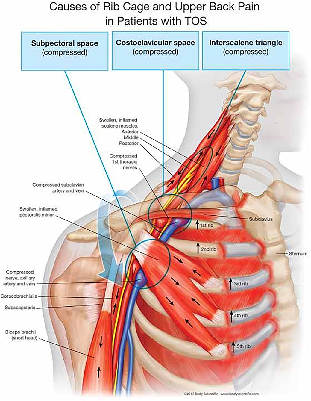
Thoracic outlet syndrome (TOS) refers to compression of the neurovascular structures passing through the thoracic outlet. It causes pain, numbness, and weakness in the arm and shoulder.
There are three types of TOS:
1. Neurogenic TOS: Compression of the brachial plexus nerves. This is the most common type and causes pain, numbness and weakness in the arm and hand. It happens due to muscle spasms or a dropped first rib compressing the nerves.
2. Venous TOS: Compression of the subclavian vein. This leads to swelling in the arm from impaired blood return. It can sometimes cause thrombosis of the subclavian vein. Trauma or anatomical abnormalities like a cervical rib are often responsible.
3. Arterial TOS: Compression of the subclavian artery. This is the least common but most serious type. It can lead to compromised blood flow in the arm. It requires prompt treatment to prevent permanent damage. An anomalous first rib or cervical rib are usually to blame.
The symptoms of TOS may include:
• Pain and numbness in the neck, shoulders, and arm. Usually aggravated by arm activity.
• Muscle weakness and fatigue in the affected arm.
• Swelling of the arm and hand. Visible veins in the neck and chest area.
• Coldness in the hands and fingers. Pale or bluish discoloration of the arm.
• Tenderness over the cervical spine, shoulder, and first rib. Trigger points may be present.
• Diagnosis is made based on symptoms, physical exam, imaging and nerve conduction studies. Chest x-ray can detect any bony abnormalities.
Treatment options for TOS include:
• Rest, immobilization, and physical therapy to relieve pressure. This is usually done for neurogenic TOS. Surgery is reserved for failed conservative treatment.
• Venous TOS may require thrombolysis, blood thinners, and correction of any anatomical cause. Surgery to resection anomalous ribs may be recommended in some cases.
• Arterial TOS often requires immediate surgical decompression by resection of anomalous ribs or muscles to restore blood flow and prevent permanent damage.
• Surgical procedures include first rib resection, anterior scalenectomy, and neurolysis of brachial plexus. They are very effective at relieving symptoms when conservative options fail or in arterial TOS.
.